Many advancements in modern medicine rely on diagnostic imaging techniques like CT scans to provide accurate and timely diagnoses. At Houston Family Doctors, we understand the significance of these imaging tools and aim to offer a comprehensive guide to understanding the basic principles of CT scans. From the non-invasive nature of the procedure to the intricate working principles of the technology involved, this guide explores the crucial components, types of scans, benefits, limitations, preparation, and what to expect during a CT scan. Stay informed and empowered when it comes to your healthcare decisions with our detailed insights into CT scans.
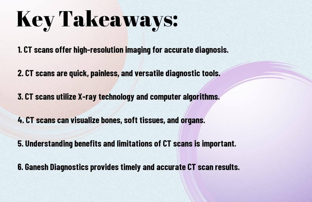
Key Takeaways:
- CT Scan Basics: CT scans are non-invasive imaging techniques that provide detailed 3D images of the body, helping physicians visualize internal structures with exceptional clarity.
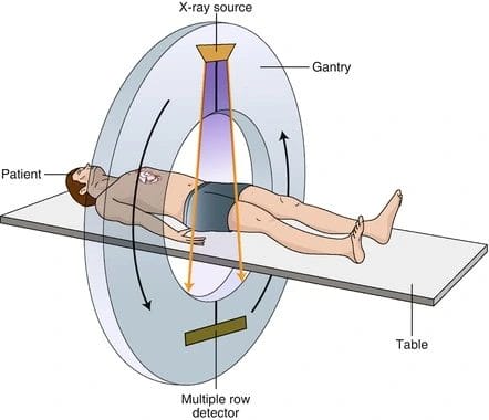
- Working Principle: CT scans use X-ray technology and computer algorithms to capture multiple X-ray projections from different angles, which are then reconstructed into cross-sectional images.
- Components of a CT Scanner: A CT scanner consists of key components like the X-ray tube, detectors, gantry, and computer system working together to produce detailed images for accurate interpretation.
- Types of CT scans: CT scans can be performed on various body parts such as the head, chest, abdomen, and pelvis to diagnose conditions like brain tumors, lung issues, and abdominal trauma.
- Benefits and Limitations: CT scans offer detailed imaging, quick and painless procedures, versatility in diagnostic applications, but come with considerations like radiation exposure, soft tissue limitations, and contrast agent risks.
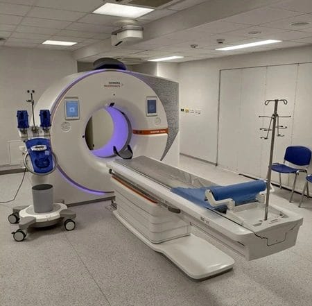
Basics of CT Scan Technology
Definition and Purpose
Definition: A CT scan, or computed tomography, is a non-invasive medical imaging technique that combines X-ray technology and computer algorithms to produce detailed cross-sectional images of the body. The purpose of a CT scan is to provide 3D representations of internal structures, enabling physicians to visualize bones, soft tissues, and organs with exceptional clarity, aiding in accurate diagnosis and treatment planning.
How CT scans Differ from Other Imaging Techniques
Other: CT scans differ from traditional X-rays by providing higher resolution, 3D images that can capture a more comprehensive view of the patient’s anatomy, including soft tissues and organs. While X-rays are suitable for imaging dense structures like bones, CT scans offer enhanced visualization capabilities that allow for a detailed examination of various body parts, making them a valuable tool in modern medicine.
The CT Scanning Process
Pre-Scan Preparations
To ensure the accuracy of the CT scan results, specific preparations may be necessary before the scan. Any fasting requirements will be communicated beforehand, especially for abdominal or pelvic scans, which may require you to fast to optimize visualization. Inform the radiologist about any medications you are taking that may affect the scan results. It’s crucial to communicate any allergies, especially to contrast agents, and disclose any underlying medical conditions that may impact the scan.
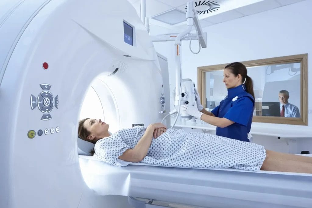
Step-by-Step Procedure During the Scan
During the CT scan, a series of steps are followed to ensure precise imaging. A table below outlines the key components and processes involved in a CT scan.
| Procedure | Description |
| Preparation | The patient is positioned on the CT scanning table. |
| Contrast Agent | If necessary, contrast agents may be administered for enhanced imaging. |
| Image Acquisition | The CT scanner captures multiple X-ray projections from different angles. |
| Communication | The radiology technologist provides instructions and ensures patient comfort. |
A CT scan is a vital diagnostic tool that provides detailed cross-sectional images, offering valuable insights for healthcare professionals. Understanding the pre-scan preparations and the step-by-step procedure during the scan can help patients feel more informed and at ease during the imaging process.
CT Scan Types and Applications
Despite the principles common to all CT scans, different types of CT scans cater to specific areas of the body and medical conditions. Understanding the variations in CT scan types is crucial for accurate diagnosis and treatment planning.
| Head CT scan | Used for visualizing the brain, skull, and associated structures. Valuable in diagnosing brain tumors, hemorrhages, and skull fractures. |
| Chest CT Scan | Provides detailed imaging of the lungs, heart, blood vessels, and surrounding structures. Useful for evaluating lung conditions and cardiovascular diseases. |
| Abdominal CT Scan | Instrumental in assessing organs and structures in the abdomen and pelvis, aiding in the diagnosis of tumors, infections, and abdominal trauma. |
Head, Chest, and Abdominal Scans
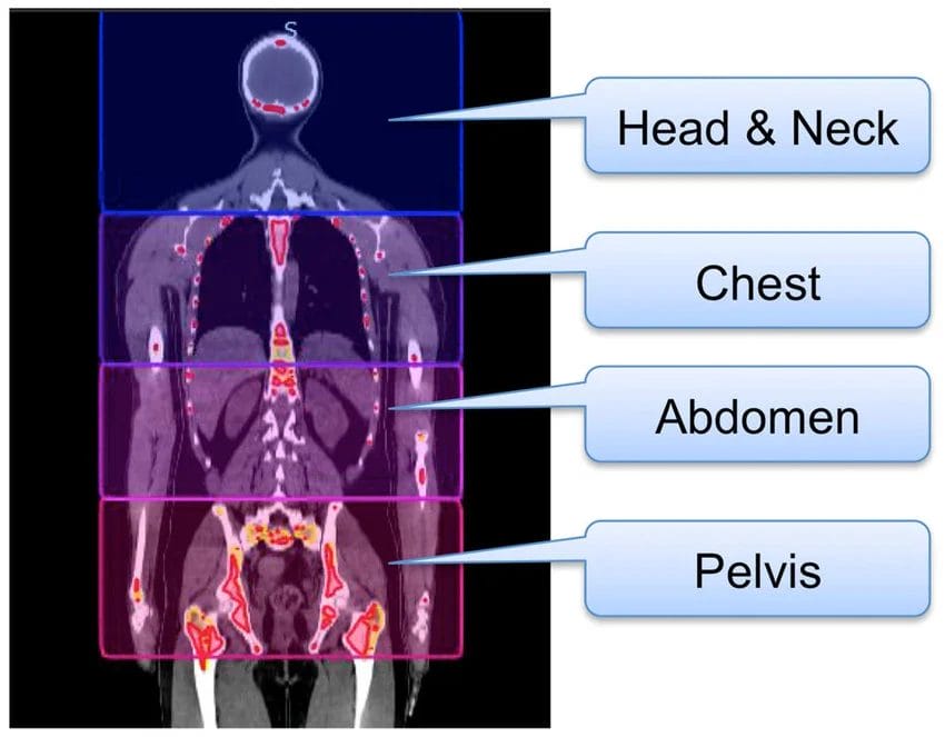
Scans targeting the head, chest, and abdominal regions offer a comprehensive view of vital structures and organs. By capturing detailed cross-sectional images, these scans assist in diagnosing a range of conditions, from brain tumors to lung abnormalities and abdominal pathologies.
Specialized CT scans for Diagnosis
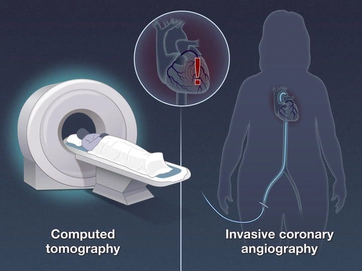
To further enhance diagnostic insights, specialized CT scans tailored to specific medical needs are available. These scans, such as angiography or cardiac CT, provide detailed images of blood vessels or heart structures, aiding in the accurate diagnosis of cardiovascular conditions.
Safety, Risks, and Considerations
Managing Radiation Exposure
Your safety is our top priority when it comes to CT scans. A necessary consideration in this regard is managing radiation exposure. While CT scans involve exposure to ionizing radiation, modern scanners utilize lower radiation doses to minimize risks. It is crucial to balance the benefits of the scan with the potential risks, especially in cases where repeated or prolonged exposure is necessary. Our team follows strict protocols to ensure the highest level of safety for all patients, prioritizing your well-being during the imaging process.
Contrast Agents and Possible Reactions
Possible reactions to contrast agents used during CT scans are rare but important to be aware of. These contrast agents are sometimes administered to enhance the visibility of specific structures or organs in the images, aiding in diagnosis. While allergic reactions or side effects are uncommon, it’s vital to inform the healthcare provider of any known allergies, especially to iodine or contrast agents. Additionally, existing medical conditions such as kidney disease or diabetes may impact the use of contrast agents. Our experienced technicians are well-equipped to handle any potential reactions and ensure your safety throughout the scanning process.
Interpreting CT Scan Results
Role of the Radiologist
For a comprehensive guide to understanding CT scan results, the role of the radiologist is pivotal. A vital member of the healthcare team, the radiologist is responsible for interpreting the images generated during the CT scan. With their expertise and specialized training, radiologists can identify abnormalities, provide accurate diagnoses, and offer valuable insights crucial for effective treatment planning.
Understanding Your Report
On receiving your CT scan report, it’s important to understand the information it contains. The report will typically detail the findings of the scan, highlighting any areas of concern or abnormalities detected. It may include descriptions of specific structures, organs, or tissues visualized during the scan, along with observations regarding their health status. Additionally, the report may offer recommendations for further diagnostic tests or consultations based on the findings.
Interpreting your CT scan report requires attention to detail and may involve medical terminology. Don’t hesitate to seek clarification from your healthcare provider or radiologist to ensure a clear understanding of the results and their implications for your health.
Summing up
With this in mind, understanding the principles of CT scans is crucial for grasping the significance of this powerful diagnostic tool in modern medicine. From the detailed imaging capabilities to the working principles and components of a CT scanner, this comprehensive guide provides a thorough overview of how CT scans work and their benefits and limitations. Patients preparing for a CT scan can gain insight into what to expect during the procedure and the importance of timely reporting in treatment planning. With a focus on patient safety, advanced technology, and expert radiologists, Ganesh Diagnostics emerges as a premier choice for all CT scan needs. Trust in their commitment to accurate and timely results, supported by their comprehensive range of scans and convenient branch locations, for a superior imaging experience.
FAQ
Q: What is a CT scan?
A: A CT scan, also known as computed tomography, is a non-invasive medical imaging technique that combines X-ray technology and computer algorithms to generate detailed cross-sectional images of the body.
Q: How does a CT Scan work?
A: The working principle of a CT scan involves X-ray beams passing through the body, detected by specialized sensors, and computer algorithms reconstructing the data into detailed cross-sectional images.
Q: What are the benefits of CT scans?
A: CT scans offer advantages such as detailed imaging, quick and painless procedures, versatility in imaging various body parts, and the option to use contrast agents for enhanced visualization.
Q: What are the limitations of CT scans?
A: Limitations of CT scans include radiation exposure, limited soft tissue differentiation, risks associated with contrast agents, and the unsuitability for pregnant women due to potential radiation risks to the fetus.
Q: How should one prepare for a CT scan?
A: Preparation for a CT scan may involve fasting for abdominal scans, adjusting medications, disclosing allergies and medical conditions, wearing appropriate clothing, and following specific instructions provided by healthcare providers for optimal results.